Home › Forums › Main Forum › Experts Corner › Fibrosis and Cirrhosis › Fibrosis Cirrhosis kPa and F Score and Hepa Score
- This topic has 9 replies, 7 voices, and was last updated 9 years, 8 months ago by
 sabrecat.
-
AuthorPosts
-
20 September 2015 at 10:27 am #1381
With Hep C treatment a key determinant of treatment outcome is liver fibrosis. Less is definitely more.
Measurement of the amount of fibrosis is called staging. There are five stages:
- F0: no scarring (no fibrosis);
- F1: minimal scarring;
- F2: scarring has occurred and extends outside the liver area (significant fibrosis);
- F3: fibrosis spreading and forming bridges with other fibrotic liver areas (severe fibrosis);
- F4: cirrhosis or advanced scarring.
In the studies patients are divided into 2 groups – have cirrhosis, don’t have cirrhosis. This is actually not realistic because you start with no fibrosis, get more and more fibrosis along the way and end up with cirrhosis.
Most HCV patients, if untreated, are expected to develop cirrhosis at about 65 years, irrespective of the age at infection. Thus, age itself seems even more important than age at infection for predicting the occurrence of liver cirrhosis.
Progression to Cirrhosis in Hepatitis C Patients: An Age-dependent Process
If you have cirrhosis or even perhaps are simply very close to having it consideration needs to be given to extending treatment from 12->24 weeks and adding Ribavirin.
Unless a liver biopsy is performed we can’t measure fibrosis directly. Instead we look to the elasticity of the liver and report the results in terms of pressures (kPA). You can see how a kPa value correlates to F

You will find more details on this at http://hepatitiscnewdrugresearch.com/fibroscan-results-the-scoring-card.html
The Hepa Score is described here: http://www.jcdr.net/articles/PDF/1136/1444_2..pdf
A hepascore value ≥ 0.50 indicates significant liver fibrosis , whereas if the result is < 0.50 significant fibrosis is absent. If the value is ≥ 0.84 , cirrhosis of the liver is likely present and if the value is < 0.84 cirrhosis is absent
YMMV
20 September 2015 at 12:29 pm #1384Ya, that’s exactly the chart my doctor here in Bangkok showed me.
12 weeks and no Ribavirin ….. YAY!
30 September 2015 at 7:55 am #1574I had a liver biopsy 10 years ago and think I was an F3
I remember there were around 4 numbers and i was shocked to see I was 1 below cirrhosisWill a fibroscan suffice to determine treatment
Or do i need to either get a new biopsy
Or get the results of the 10 year old biopsy
52 y.o. G3a for about 30 years
Previous tx 2004 interferon/ ribavarin
2004: ALT 624 AST 263Pre tx test 23/10/15: ALT 153 AST 128 VL 11 849 493
6/11/15: Sof/ dac started
26/11/15: ALT 41 AST 41
7/12/15: ALT 36 AST 30 Virus undetected2004 biopsy F3
Fibroscan appt Jan 11 2016.11 June 2016 at 4:05 am #18866My AST/ALT score is F1 and my APRI score is F4?
Noticed in one article: PWE-121 Comparison Of Kings Score, Apri and AST/ALT Ratio in Determining Severity of Liver Disease Versus Liver Biopsy in Gut 2013;62
“Conclusion Conclusions The AST/ALT ratio had the greatest diagnostic accuracy in determining significant fibrosis or cirrhosis. The Kings score performed better than APRI. AST/ALT is a simple guide to determine significant fibrosis and cirrhosis in liver disease.”
While I appreciate that any F score above 0 means some scaring, why the difference?
Note: This posting in no way supports or promotes the procurement of, or even the mention of, generic products, brand substitution, or even talking to a pharmacist or doctor about medications that one is to take. If anyone reading this post suspects that this posting has directly, or indirectly, led the reader to contemplate generic medications, or thinking for themselves, then they should report this post to the TGA – attention: ‘The Boofhead in charge of wasting tax payer’s money for no good reason’.
11 June 2016 at 11:16 am #18910My AST/ALT score is F1 and my APRI score is F4?
Hey Sabrecat, Mine too!
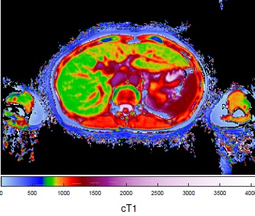
Had the amazing new LiverMultiScan and I was overall F1 with small ‘spots’ of higher fibrosis in the center & left lobe area (F3/4?) , but not joining up, just small spots. However, instinct tells me small spots can cause some problems, they seem to be where there are other organs eg heart and a lot going on in that area, maybe easier for the virus to hide in? and harder for the meds to reach possibly? Again, just my instinct. I can see why exercise is helpful, looking at my LIF scan. One thing I really learned after this new scan, is fibrosis is not necessarily evenly spread.
Added LIF image below as my blog is long and it’s hard to find; this is a mix of 4 scans :
ps green is good, red is bad

GT1a Dec14 F2/8.7 VL 900000-2.5M
Jan16 Hepcivir-L MonkMed/Redemption
Baseline: VL 913575 Alt 76 Platelets low
Wk2 VL1157 Alt 23
DET Wk 8 VL 32 Alt19 ‘In the slow lane’
June16 Fibro 5.7 F0/1 LIF 1.5
Wk 11 VL<12 Alt 13 Det/Unq
Extending tx 12 wks Mylan Sofo/Dac MonkMed
Wk 14 VL <12 Det/Unq
Wk 16 VL UNDETECTED
Wk 22 + 4 Wks Sunprevir FixHepC
Wk 24 UNDETECTED Alt 13
Wk 12 post tx SVR12 Wk 26 SVR24
Thank-you Tim, Dr Debasis @ MonkMed & Dr Freeman @ Fix HepC14 June 2016 at 6:03 pm #19051Hi London Girl,
wish I had that scan thingo here 18 years ago – they stabbed me five times during a liver biopsy!
Could not get a fibroscan recently because of a previous liver resection – so I am guessing about F scores but have to accept F3 -> F4…
I am also a bit worried about ongoing scans (for HCC) that may involve contrast dyes. Necessary, but I often worry about having too much of a good thing.
yours
Jeff
P.S.
did you get around to making a T shirt with the scan on it??14 June 2016 at 9:58 pm #19058Hey Jeff, The great thing about the new LiverMultiScan is it doesn’t need contrast dye at all, they have taken this into account.
I know this is becoming more international, how long it will take to be a regular thing where they take it on I don’t know, but you can check it out here :
http://perspectum-diagnostics.com/products/livermultiscan/
Now FDA approved for the USA.LG
GT1a Dec14 F2/8.7 VL 900000-2.5M
Jan16 Hepcivir-L MonkMed/Redemption
Baseline: VL 913575 Alt 76 Platelets low
Wk2 VL1157 Alt 23
DET Wk 8 VL 32 Alt19 ‘In the slow lane’
June16 Fibro 5.7 F0/1 LIF 1.5
Wk 11 VL<12 Alt 13 Det/Unq
Extending tx 12 wks Mylan Sofo/Dac MonkMed
Wk 14 VL <12 Det/Unq
Wk 16 VL UNDETECTED
Wk 22 + 4 Wks Sunprevir FixHepC
Wk 24 UNDETECTED Alt 13
Wk 12 post tx SVR12 Wk 26 SVR24
Thank-you Tim, Dr Debasis @ MonkMed & Dr Freeman @ Fix HepC15 June 2016 at 1:48 am #19063Hi Sabre
I made LG a t shirt and flares ages ago
They’re on her blog and on mine
Ariel
Here are screenshots of what you missed for a laugh Attachments:15 June 2016 at 2:11 am #19064
Hi London Girl – that is an amazing MRI imaging. It looks so much more accurate than Fibroscan as it can check all areas of the liver. My lay assumption is that even if scored F1 (my case) on Fibroscan or conventional Ultra-sound, we might be missing those red (bad) spots where it is more difficult for the DAAs to reach. Cool pants by the way.


Blood transfusion in 1992 – Diagnosed in 2007
Tx naive -G1b – F1
VL 2.270.000
ALT 40
Start tx June 4th/2016 with DAAs – Sof/Led from India
Bloods on two weeks of tx (June 18th)
AST 17 – ALT 10 – GGT 19
Virus UND
Bloods on six weeks of tx (July 16th)
AST 17 – ALT 8 – GGT 12
Virus UND
EOT on August 8th (did 9 weeks and 3 days)SVR 4 Virus UND (September 7th)
AST 13 – ALT 5SVR 14 Virus UND (November 12th)
15 June 2016 at 5:44 am #19076Hi Ariel,
Was going to ask you to make some for me, but mine are in crummy black and white.
But maybe you could photoshop out the scarring so I don’t worry as much; or for some of our friends who have livers not bad enough for treatment to be covered, add some marks here and there?
yours
Jeff
-
AuthorPosts
- You must be logged in to reply to this topic.

